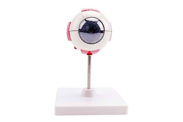

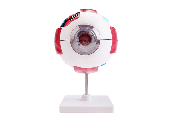


30409200301 Eye Anatomy Model
Product Description:
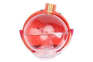
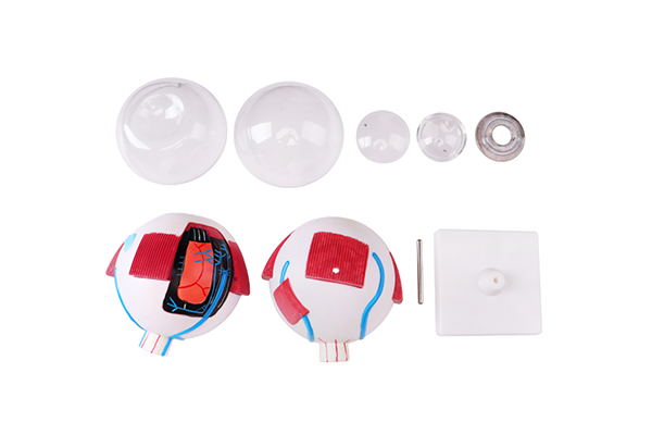
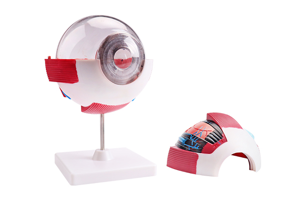
● Product: This is a six-times enlarged adult eyeball model mounted on a stand.
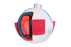
● Cutaway: The model features a median horizontal cut through the anterior and posterior poles of the eyeball, displaying the three layers of the eyeball wall, the lens, vitreous humor, and iris (all removable).
● External Structures: The external structures of the eyeball wall include the eyeball itself, cornea, sclera, iris, pupil, the cut ends of the six extraocular muscles, optic nerve, vortex veins, long posterior ciliary artery (iris artery), and short posterior ciliary artery (choroidal artery).
● Internal Structures: The internal structures of the eyeball wall and its cross-section primarily show the outer layer (the cornea at the front one-sixth and the sclera at the remaining five-sixths), the middle layer (iris, ciliary body, and choroid), and the inner layer (retina with the optic disc at the posterior part, macula, retinal vessels, lens, and vitreous humor).